Dashboard
Welcome back, Dr. Bao Yuntong • March 29, 2026
Powered by Medical Large Model v3.2 • Diagnostic Accuracy 98.5%
Total Patients
156
↑ 12% increase from last monthPending Cases
24
Please process in a timely manner ⏳AI Diagnostic Accuracy
98.5%
FDA Certified Standard ✅High-Risk Cases
8
Requires specialist review 🔴Welcome to Salus AI Intelligent Ultrasound-Assisted Diagnosis System
👨⚕️ Physician: Brittany Bao
📌 System Features: AI Ultrasound Image Analysis, Gastric/Pancreatic Lesion Diagnosis Assistance, Patient Records Management, Diagnostic Report Generation
⚠️ Important Notice: AI analysis results are for clinical reference only, final diagnosis must be confirmed by licensed physicians
Recent Patient Examination Records
| Patient ID | Patient Name | Age | Gender | Examination Type | Risk Level | Status | Action |
|---|---|---|---|---|---|---|---|
| #178 | James Anderson | 42 | Male | Gastric Ultrasound (Gastric Filling Contrast) | High Risk | Pending | View |
| #076 | Emma Wilson | 35 | Female | Pancreatic Ultrasound (Pancreatic Parenchyma Evaluation) | Medium Risk | Completed | View |
| #103 | Sofia Martinez | 28 | Female | Combined Gastric + Pancreatic Ultrasound | Normal | Completed | View |
| #045 | Michael Brown | 52 | Male | Pancreatic Contrast-Enhanced Ultrasound | Medium Risk | Pending Review | View |
System Workflow
Salus AI Complete Workflow for Gastric/Pancreatic Ultrasound Diagnosis

Gastric/Pancreatic Ultrasound Case Examples

Case 1: Routine gastric ultrasound scan with clear gastric wall layers and no obvious space-occupying lesions.
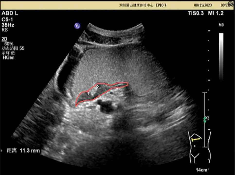
Case 2: Annotated pancreatic ultrasound image with a 11.3mm abnormal echo area marked in red, identified as high-risk lesion by AI.
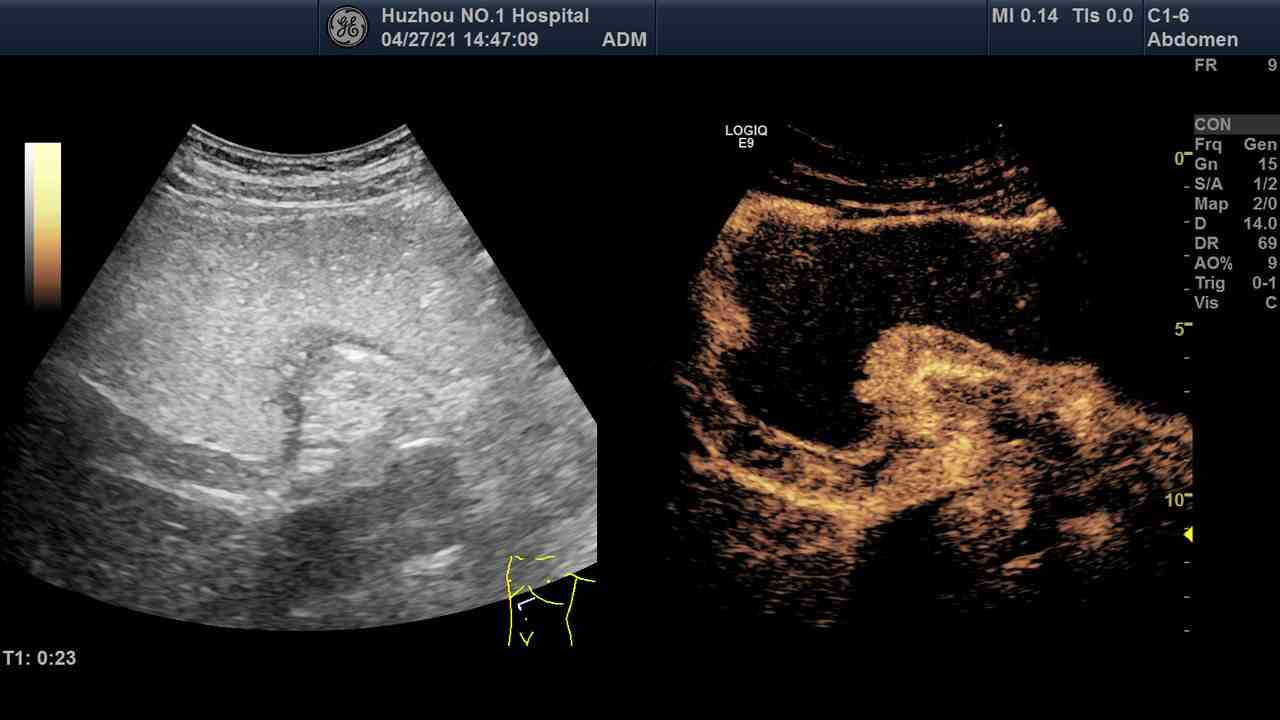
Case 3: Contrast-enhanced gastric/pancreatic ultrasound image showing conventional ultrasound on the left and contrast imaging on the right, clearly demonstrating lesion vascular characteristics.
Note: The above are clinically real gastric/pancreatic ultrasound examination cases. The AI system can automatically identify abnormal areas and label risk levels.
Gastric/Pancreatic Ultrasound Image Upload & AI Analysis
Patient Information & Image Upload
Please upload gastric/pancreatic ultrasound image
Click or drag to upload gastric/pancreatic ultrasound image/video
AI Diagnostic Analysis Results
Upload gastric/pancreatic ultrasound image and click analyze to view AI diagnostic results
Patient Records Management
All Patient Examination Records
| Patient ID | Patient Name | Age | Gender | Examination Type | Risk Level | Status | Action |
|---|---|---|---|---|---|---|---|
| No medical records available. Please upload gastric/pancreatic ultrasound images to generate medical records. | |||||||
Standard Ultrasound Diagnostic Workflow for Gastric/Pancreatic
AI-Assisted Complete Workflow for Gastric/Pancreatic Ultrasound Diagnosis
Important Notes for Gastric/Pancreatic Pathological Diagnosis
1. Patients must be fully prepared before gastric/pancreatic ultrasound examination, and contrast agent use must follow standard procedures
2. The AI system can quickly identify lesions such as gastritis, gastric ulcers, pancreatitis, pancreatic masses, with automatic marking of high-risk cases
3. All AI analysis results must be finally reviewed and confirmed by licensed physicians
4. Diagnostic reports are automatically filed and support one-click export, printing, and sharing